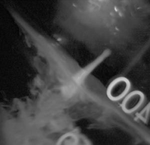
Scientists have been able to use off-the-shelf DSLR cameras and digital movie projectors to create a high speed high resolution tool capable of advancing detailed scientific imaging at consumer market prices.
See: Cameras of the future: heart researchers create revolutionary photographic technique
“Scientists funded by the Biotechnology and Biological Sciences Research Council and the British Heart Foundation at the University of Oxford have developed a revolutionary way of capturing a high-resolution still image alongside very high-speed video – a new technology that is attractive for science, industry and consumer sectors alike.”
The fact that this can be achieved with off-the-shelf existing cameras means that the technology can (and most likely will ) be incorporated into readily available cameras for all of us to use. – Yes, even in nonscientific unfunded applications, like any other scene we might shoot of friends, pets, models, etc.
“What’s new about this is that the picture and video are captured at the same time on the same sensor” said Dr Bub. “This is done by allowing the camera’s pixels to act as if they were part of tens, or even hundreds of individual cameras taking pictures in rapid succession during a single normal exposure. The trick is that the pattern of pixel exposures keeps the high resolution content of the overall image, which can then be used as-is, to form a regular high-res picture, or be decoded into a high-speed movie.”
“This could have everyday applications for everything from CCTV to sports photography and is already attracting interest from the scientific imaging sector where the ability to capture very high quality still images that correspond exactly to very high speed video is extremely desirable and currently very expensive to achieve. The technology has been patented by Isis Innovation, the University of Oxford’s technology transfer office, which provided seed funding for this development and welcomes contact from industry partners to take the technology to market. The research is published today (14 February 2010) in Nature Methods.
“Dr Peter Kohl and his team study the human heart using sophisticated imaging and computer technologies. They have previously created an animated model of the heart, which allows one to view the heart from all angles and look at all layers of the organ, from the largest structures right down to the cellular level. They do this by combining many different types of information about heart structure and function using powerful computers and advanced optical imaging tools. This requires a combination of speed and detail, which has been difficult to achieve using current photographic techniques.”
The research may soon move from the optical bench to a consumer-friendly package. Dr. Mark Pitter from the University of Nottingham is planning to compress the technology into an all-in-one sensor that could be put inside normal cameras. Dr Pitter said: “The use of a custom-built solid state sensor will allow us to design compact and simple cameras, microscopes and other optical devices that further reduce the cost and effort needed for this exciting technique. This will make it useful for a far wider range of applications, such as consumer cameras, security systems, or manufacturing control.”
For more tech on the method for multiplexing temporal information from a imaged scene into a single frame of an image, while maintaining full spatial resolution for the image, see:
Temporal Pixel Multiplexing
The image above is protected by copyright law and may be used with acknowledgement of the Nature Methods article.
This will change the way all of our digital cameras process the visual information.
More and better pix for all.
All Power to the Sensor!
This is SO high-tech for me……but cool!
It’s high-techy for almost everybody.
The coolest part is that the outcome of this research may be coming to a camera near you, and you won’t need to understand the tech behind it.